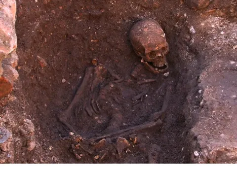
Scientists from the University of Leicester said that DNA evidence, historical records and carbon dating all suggest that this skeleton belonged to Richard III, the last King to die in battle.
How did they do it? Here’s three ways the team used the latest scientific techniques to identify the remains of the lost King.
Determining the time of death – radiocarbon dating

The first task the team set on was to establish that the skeletal remains were from the right time period. To do so the team used radiocarbon dating to determine the age of the remains. It’s a technique commonly used to estimate the age of any organic substance. It relies on the fact that all living things contain radioactive carbon isotopes obtained through their diet.
When a human dies, they contain a known concentration of carbon isotopes, but as the body decays this concentration drops. These isotopes decay at a regular rate, known as their half-life, and so by measuring the remaining radioactive carbon in the body it's possible to estimate the age of the remains.
This technique was employed to give the approximate date of death as lying between AD1475-1530 (with a 69% confidence). Current knowledge suggests that Richard III died in 1485.
Determining the cause of death–micro-CT scanning

Once it was confirmed that the time of death fit what historians know about Richard III’s demise, scientists began to reconstruct the skeleton digitally.
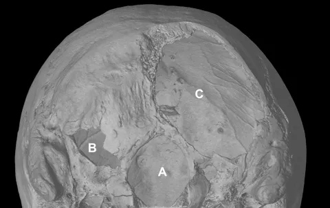
Micro-computer X-ray tomography (micro-CT) was used to build a 3D profile of the skeleton and investigate potentially fatal injuries to the skull in as much detail as possible.In micro-CT, an X- ray emitter generates electrons, which are fired towards the target material – in this case the skull. When they strike, they slow down, generating X-ray emissions, which are picked up by an X-ray detector. This all happens while the sample is rotated to create a fully-detailed 3D image of the skull.
The 3D model permits researchers to study the remains in greater detail with no risk of harming the fragile bones. In particular, the visible wounds and the extent of scoliosis – curvature of the spine, which Richard III is famed for.
Applying the micro-CT technique to the bones allowed the team to study them in great detail, in particular the visible wounds and the extent of the scoliosis of the spine.
By analysing the scans, the archeologists discovered that the back of Richard III’s skull had been sliced by a halberd (a type of medieval axe) and that he’d been stabbed in the cheek, resulting in facial fractures and missing portions of his skull.
Determining the identity–extraction and analysis of DNA

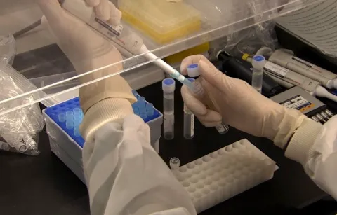
Mitochondrial DNA testing was used to determine whether the remains irrefutably belonged to Richard III.
Mitochondria are small parts of the cell whose function it is to convert chemical energy (from food) into a form that the body can use. Unlike nuclear DNA (nDNA), which is inherited half from the mother and half from the father, mtDNA is passed down intact from the mother alone. This is because mitochondria in sperm cells are destroyed as part of the fertilisation process. As result, if a female line of the family remains unbroken the mtDNA remains largely the same, allowing scientists to trace this lineage back hundreds of years from the present day.
Fortunately, due to their preservation, the teeth yielded usable mtDNA for analysis. The DNA was then compared and matched to two living ancestors of the King who were traced down through an all-female line of descent by genealogists.
Follow Science Focus onTwitter,Facebook, Instagramand Flipboard