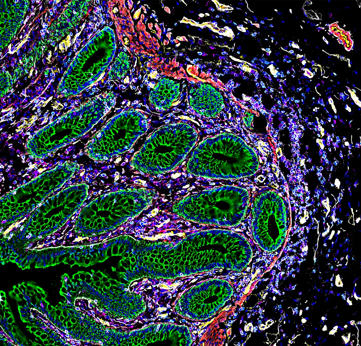
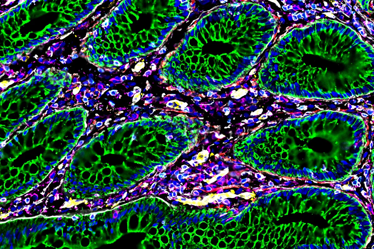
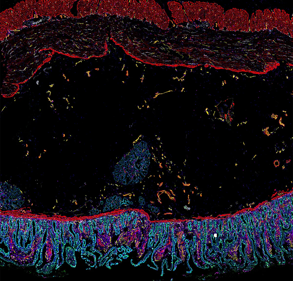
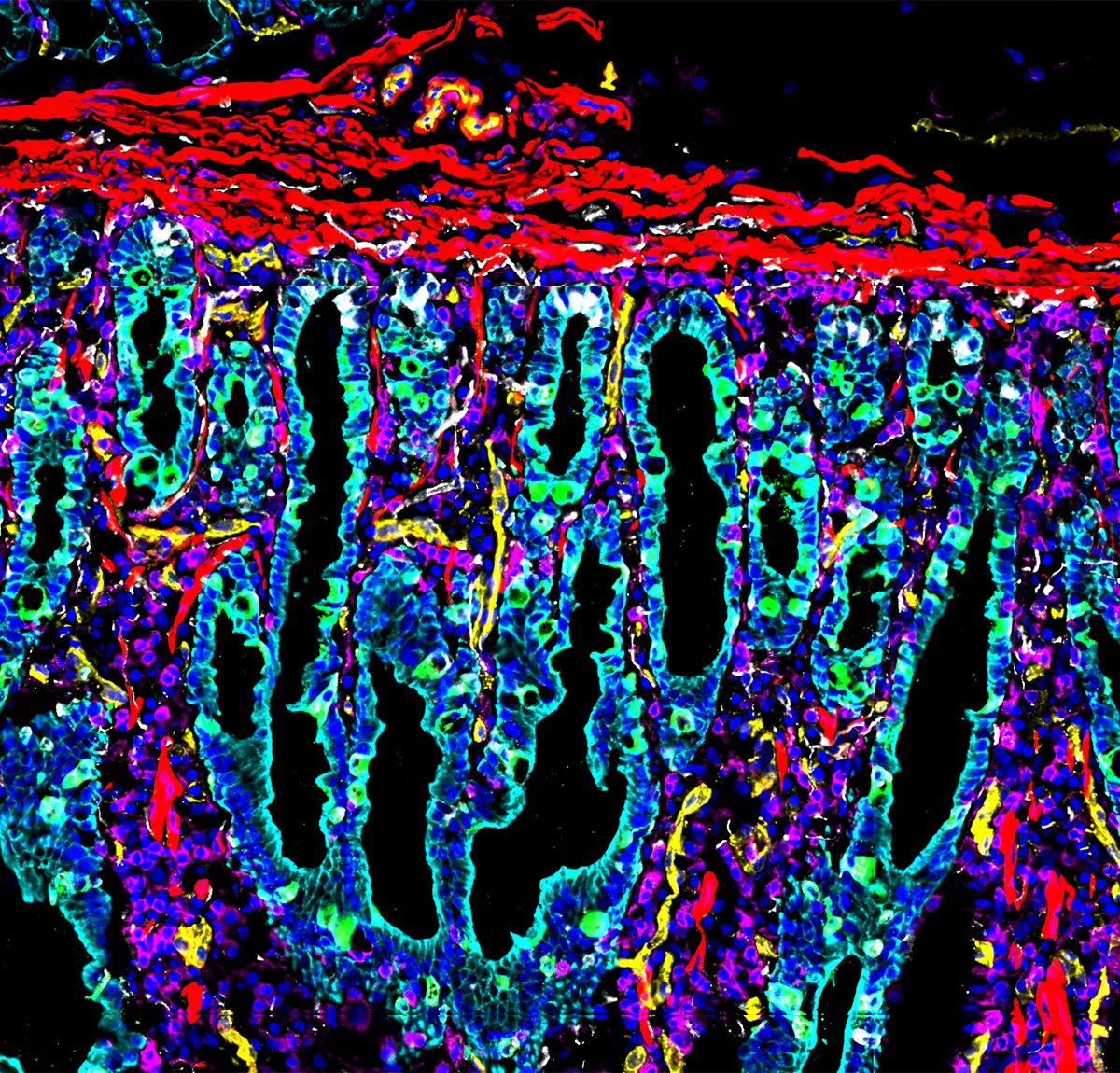
Researchers from Stanford Medicine in the USA have used an advanced new imaging technique to map the human digestive system. This revealed that our gut is like other neighbourhood communities, with different types of cells cooperating with each other to digest food and fight off bugs and infections.
Scientists examined eight regions of the small and large intestines of nine deceased individuals. To image these regions they used a technology called 'co-detection by indexing' (CODEX). This involves staining and washing the gut tissue repeatedly with fluorescent antibodies which bind to specific proteins, and thus enable easier imaging.
By combining CODEX as well as other new imaging and sequencing technologies, the researchers were able to map these neighbourhoods down to the level of individual cells – something that has never been achieved before.
Once the gut had been mapped, the researchers were able to identify 20 distinct cellular neighbourhoods within the human digestive system. These include epithelial cells that make up the intestinal lining, connective tissue cells, nerve cells and immune cells, as well as blood vessels.


The researchers hope that these images will be used in a clinical setting to help diagnose conditions such as irritable bowel syndrome and early-stage colon cancer, where healthy and unhealthy digestive system images can be compared and analysed by doctors.
Eventually, the team also aims to produce a three-dimensional map of the gut to better understand the nerve structures and blood vessels of the digestive system, which in turn will help with diagnosis and treatments.
Read more:
