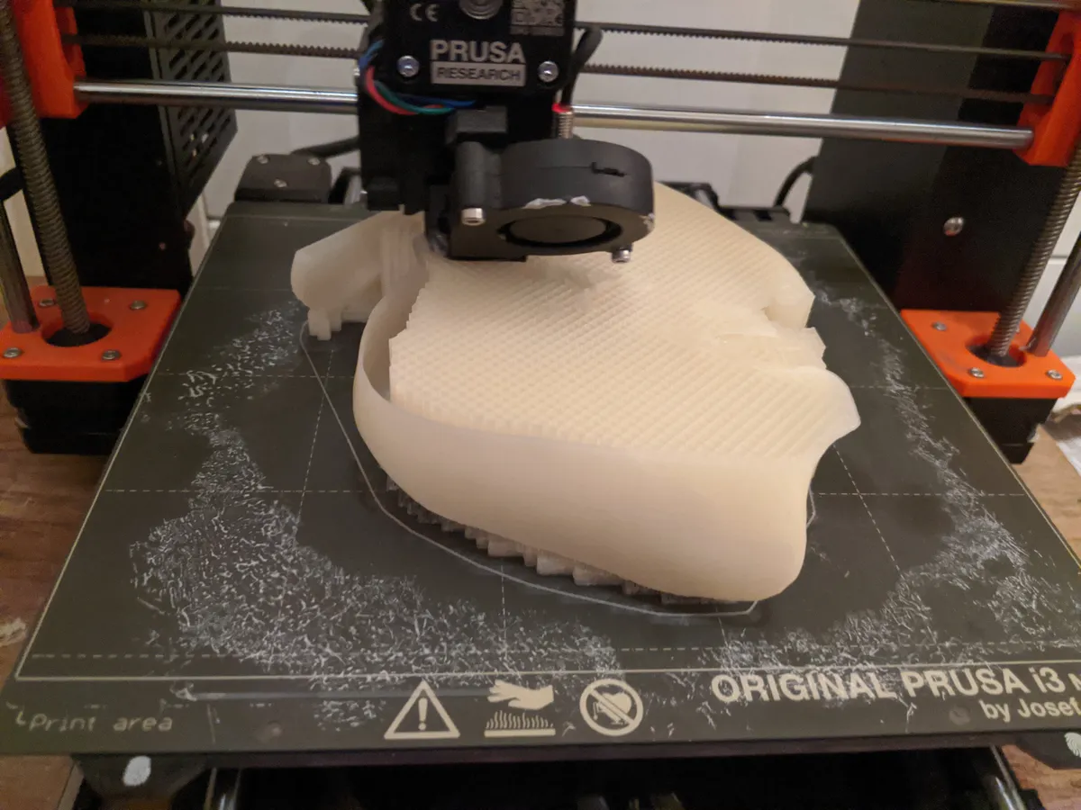
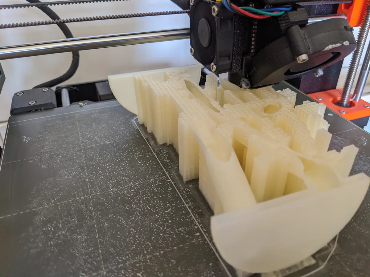
Scientists at Nottingham Trent University have developed a method of using scans of cancer patients’ organs to 3D print realistic models so that surgeons can rehearse procedures before they take place in real life. The technique recreates the individuality of a patient’s organ, with blood vessels, tissue and any tumours all being included in the completed model.

The model liver was constructed using existing 3D scan data taken from an anonymous cancer patient.

The team used a combination of synthetic fibres and gels to 3D print an accurate model of the organ, layer by layer.

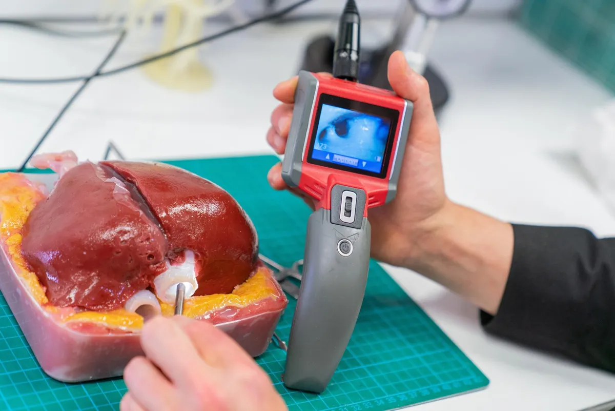
The completed liver model has a texture similar to that of a real diseased organ, including different tissue hardnesses in the blood vessels, liver tissue and the tumour itself. It is even filled with imitation blood, to make it as realistic as possible and could even provide trainee surgeons with the opportunity to learn how to remove tumours.
As the process accurately recreates an organ’s internal structure, the model allows surgeons to use real tools to practise complicated procedures such as endoscopies.

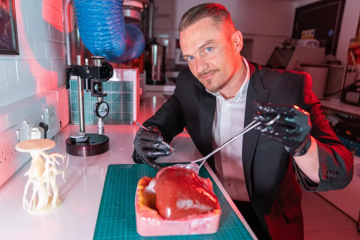
Senior research fellow Richard Arm demonstrates how a surgeon might practise an operation on the 3D-printed organ prior to performing the surgery on a living patient.
“Every patient is unique and has organs of different shapes, sizes and constructs, so there can be many hidden complications that they [the surgeons] have to deal with,” he said. “But this research shows how existing scan data and modern 3D print processing methods can dramatically improve the preparation available before the first incision is even made.”
Read more about 3D printing: