Hidden away in the dusty vaults of natural history museums worldwide is a treasure trove of rare species, collected over hundreds of years and out of public view. But now, thanks to a new project, over 13,000 specimens will be available for anyone to access digitally.
Collated over the last five years, the openVertebrate (oVert) collaboration between 18 institutions has produced an amazing collection of 3D reconstructions of vertebrate specimens.
This involved oVert project members taking thousands of CT scans (a type of X-ray image) of vertebrate species found in their collections, including many examples of amphibians, reptiles, fish and mammals. Even a humpback whale was painstakingly scanned to produce an impressive 3D model.
This new approach will open up a whole world of research possibilities for scientists and researchers, as well as benefitting educators and artists who can use these resources to illustrate accurate models of a huge range of species.
Scientists have already used data from the oVert project to gain astonishing insights into the natural world, and as technology becomes evermore advanced this resource will become even more essential.
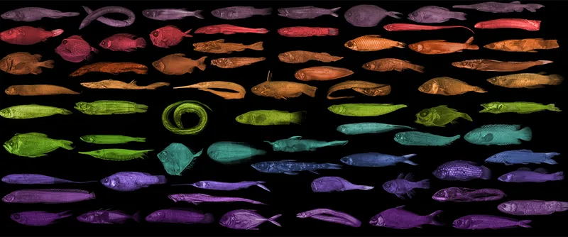
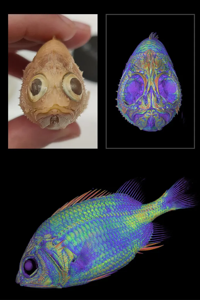
Fish varieties
Specimens (like these different species of fish) once restricted solely to the scientists who study them are now available as 3D models to everyone. Photo by openVertebrate
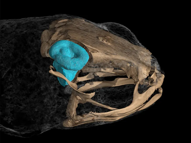
The vestibular system in frogs
The vestibular system in pumpkin toadlets (Brachycephalus) is the smallest ever observed in vertebrates. Still, it takes up proportionally more space within their heads than in larger organisms, leaving them unable to balance while jumping. Photo by openVertebrate
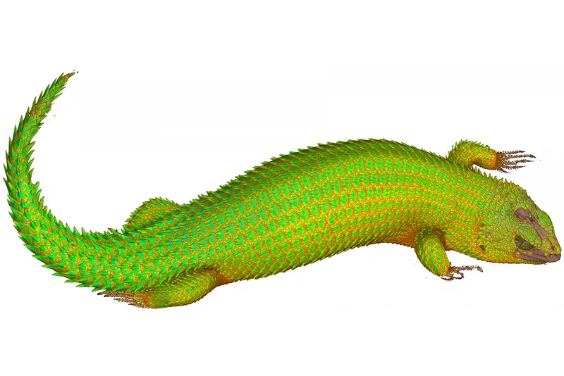
Animal scans with incredible detail
A scan of Hosmer's spiny-tailed skink (Egernia hosmeri) demonstrates the level of detail these animal scans can reveal. Photo by openVertebrate
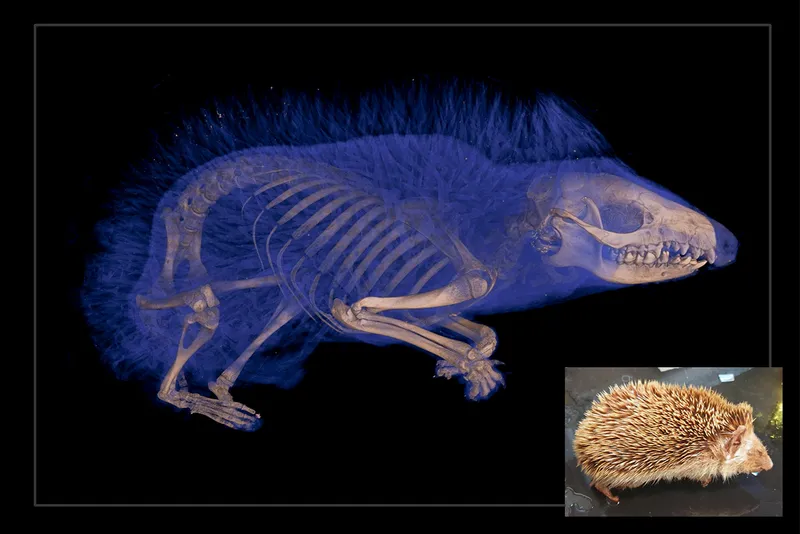
The structures hidden within
With CT scanning, scientists can study a specimen's internal anatomy without the need for dissection – this hedgehog image being a good example. Photo by openVertebrate
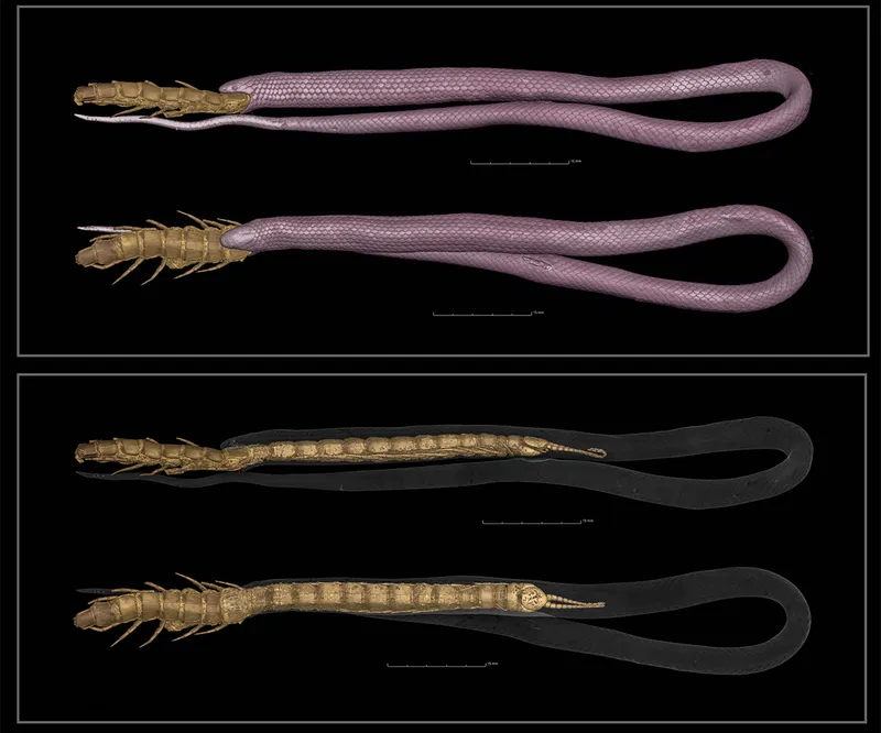
A centipede too far
Researchers using the oVert system were able to perform a digital dissection of a rim rock crowned snake (Tantilla political) – North America's rarest snake. This particular specimen had died while trying to eat a centipede. Photo by openVertebrate
Unprecedented diversity
The primary goal behind the oVert project is to image as great amount of diversity across the vertebrate tree of life as possible, including fish, reptiles and mammals. Photo by openVertebrate
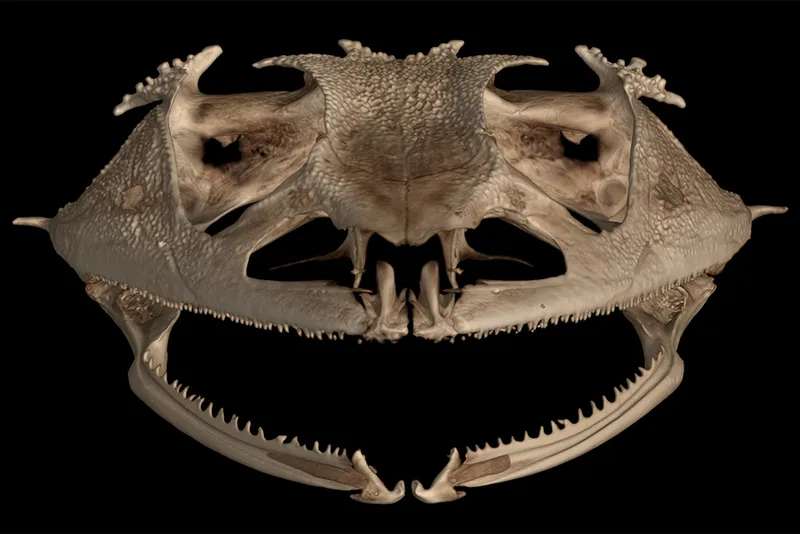
Evolutionary insights
An analysis of oVert specimens revealed that frogs have lost their teeth over 20 times throughout their evolutionary history, more than any other vertebrate group. Photo by openVertebrate
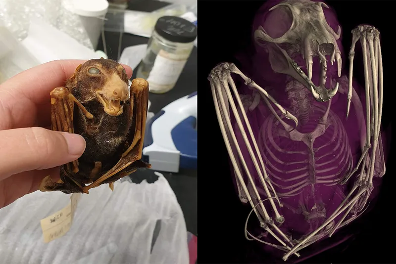
A digital museum
On the left, a scientist holds a black-bellied fruit bat (Melonycteris melanops). The image on the right shows the same bat after it was scanned in 3D. Photo by openVertebrate
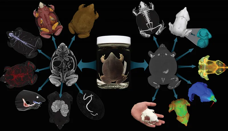
A frog flowchart
Using various methods, researchers can reconstruct museum specimens as digital 3D models. Photo by openVertebrate
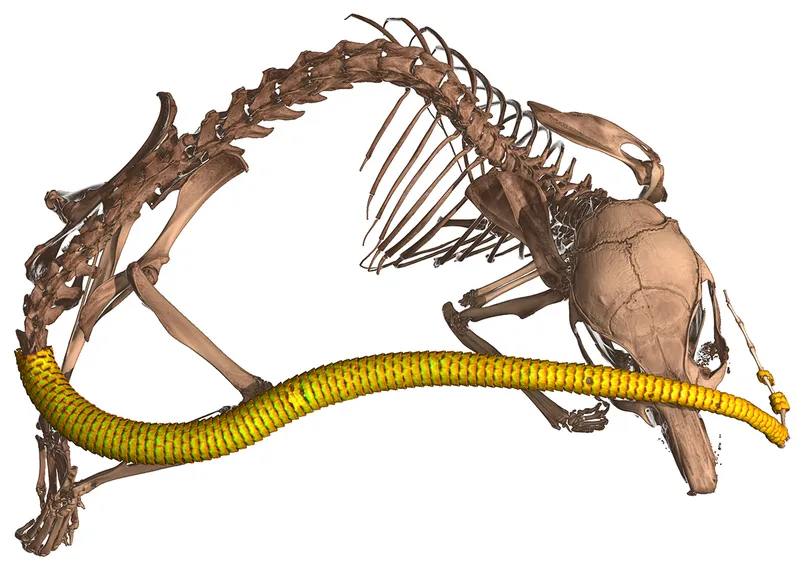
Osteoderms in spiny mice
One of the researchers was conducting routine CT scans of spiny mice and was surprised to find their tails were covered with an internal coat of bony plates, called osteoderms. Before this discovery, armadillos were considered to be the only living mammals with these structures. Photo by openVertebrate
James Cutmore is the picture editor of BBC Science Focus Magazine. He has worked on the magazine and website for over a decade, telling compelling science stories through the use of striking imagery. He holds a degree in Fine Art, and has been nominated for the British Society of Magazine Editors Talent Awards, being highly commended in 2020. His main areas of interest include photography that highlights positive technology and the natural world. For many years he was a judge for the Wellcome Trust's image competition, as well as judging for the Royal Photographic Society.
This website is owned and published by Our Media Ltd. www.ourmedia.co.uk