When you look at around you there so is much more to see than just what your eyes can pick out. Just out of sight is a tiny, microscopic world teeming with life, from microbial beings to intricate patterns formed in everyday materials that can both delight and inform, for example Jean-Marc Babalian’s photograph of Volvox algae is reminiscent of Pac Man, or the Netherlands Cancer Institute’s of human skin cells helping us understand how diseases progress. Both of these photos were at the top of theNikon Small Worldcompetition, which rewards those photographers putting their lenses closer than any of us could dream with our regular cameras.
20th Place - Tracy Scott
Ithaca, New York, USA
Aspergillus flavus (fungus) and yeast colony from soil
Transmitted Light, 40x
19th Place - Dr. Dylan Burnette
Vanderbilt University School of Medicine, Department of Cell and Developmental Biology
Nashville, Tennessee, USA
Embryonic body wall from a developing Mus musculus (mouse)
100x (objective lens magnification)

18th Place - Christian Gautier
Biosphoto
Le Mans, France
Synapta (sea-cucumber) skin
Polarised Light, 100x
17th Place - Harald K. Andersen
Steinberg, Norway
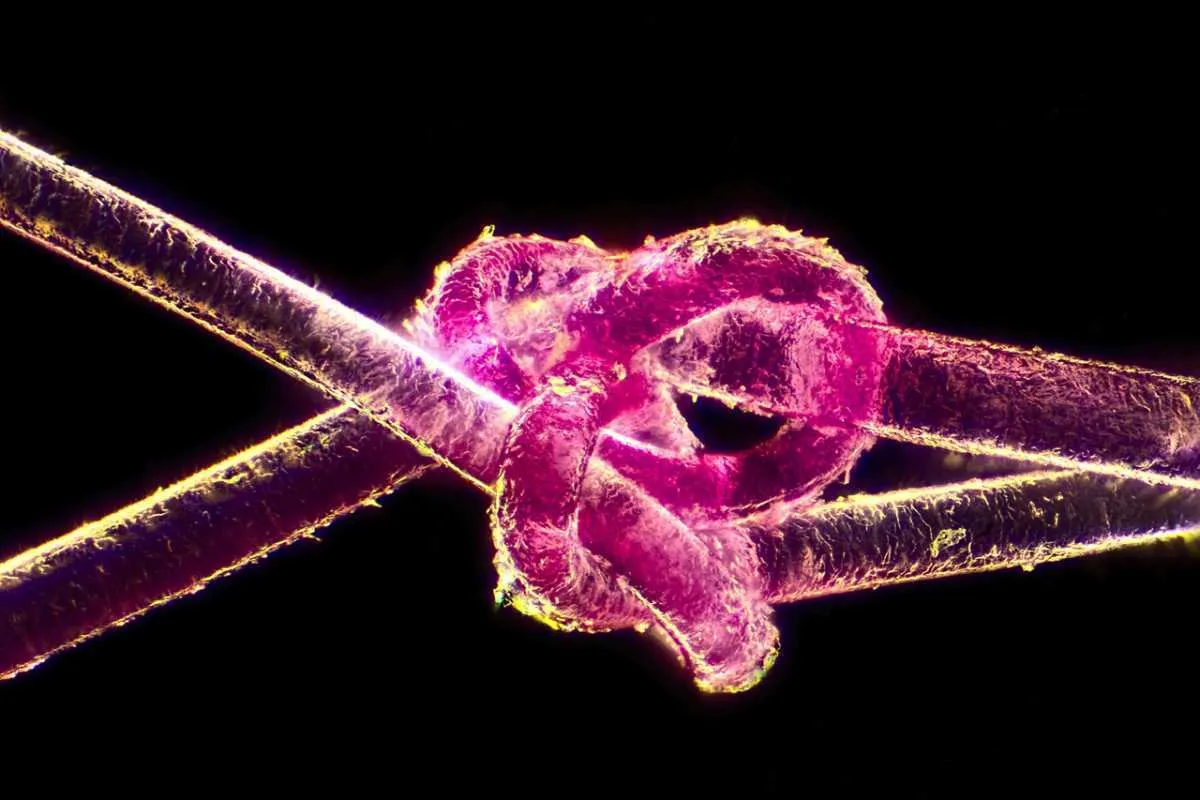
Dyed human hair
Darkfield, 40x
16th Place - Marek Miś
Marek Miś Photography
Suwalki, Poland
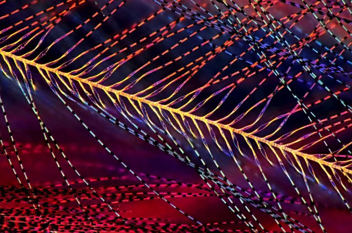
Parus major (titmouse) down feather
Polarised Light, Darkfield, 25x

15th Place - Dr. Rick Adams
University of Northern Colorado, Department of Biological Sciences
Greeley, Colorado, USA
3rd trimester fetus of Megachiroptera (fruit bat)
Darkfield, Stereomicroscopy, 18x
14th Place - David Millard
Austin, Texas, USA
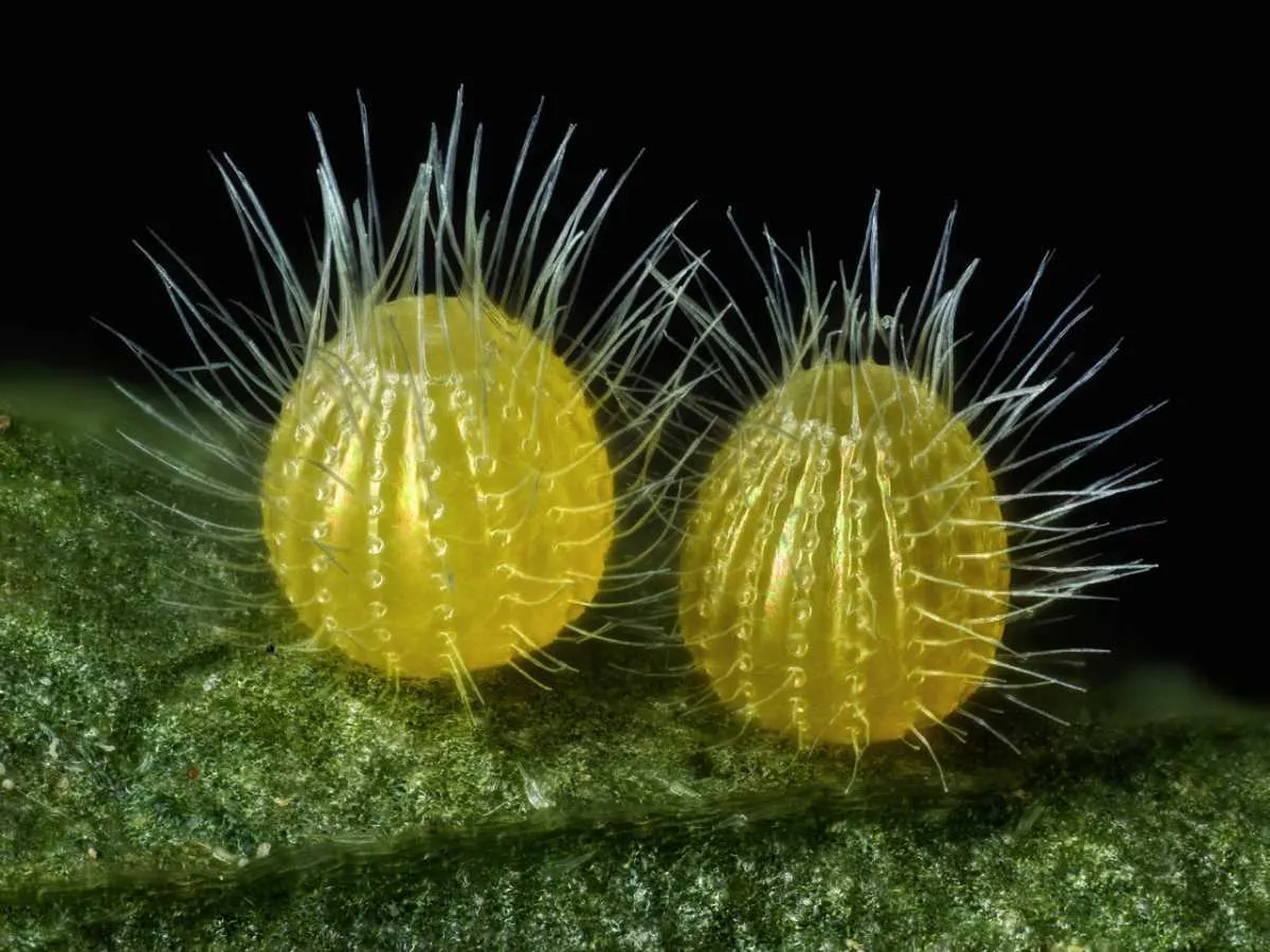
Common Mestra butterfly (Mestra amymone) eggs, laid on a leaf of Tragia sp. (Noseburn plant)
Incident Illumination, Image Stacking, 7.5x (objective lens magnification)
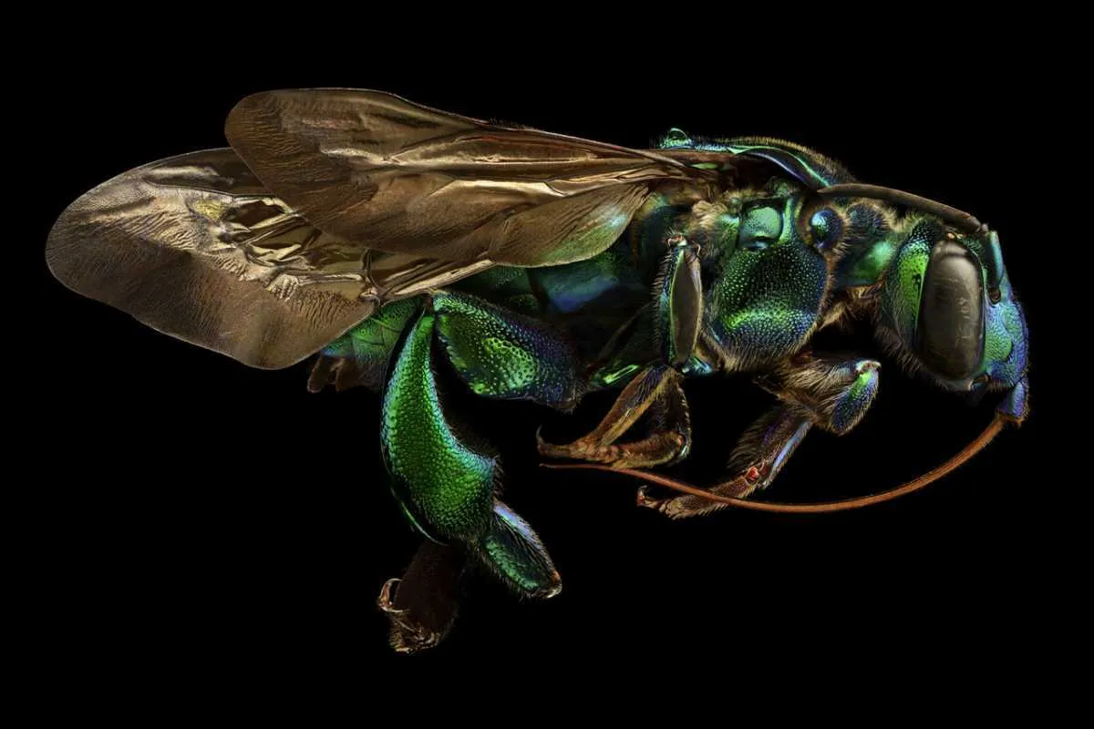
13th Place - Levon Biss
Levon Biss Photography Ltd
Ramsbury, United Kingdom
Exaerete frontalis (orchid cuckoo bee) from the collections of the Oxford University Museum of Natural History
Reflected Light, 10x (objective lens magnification)
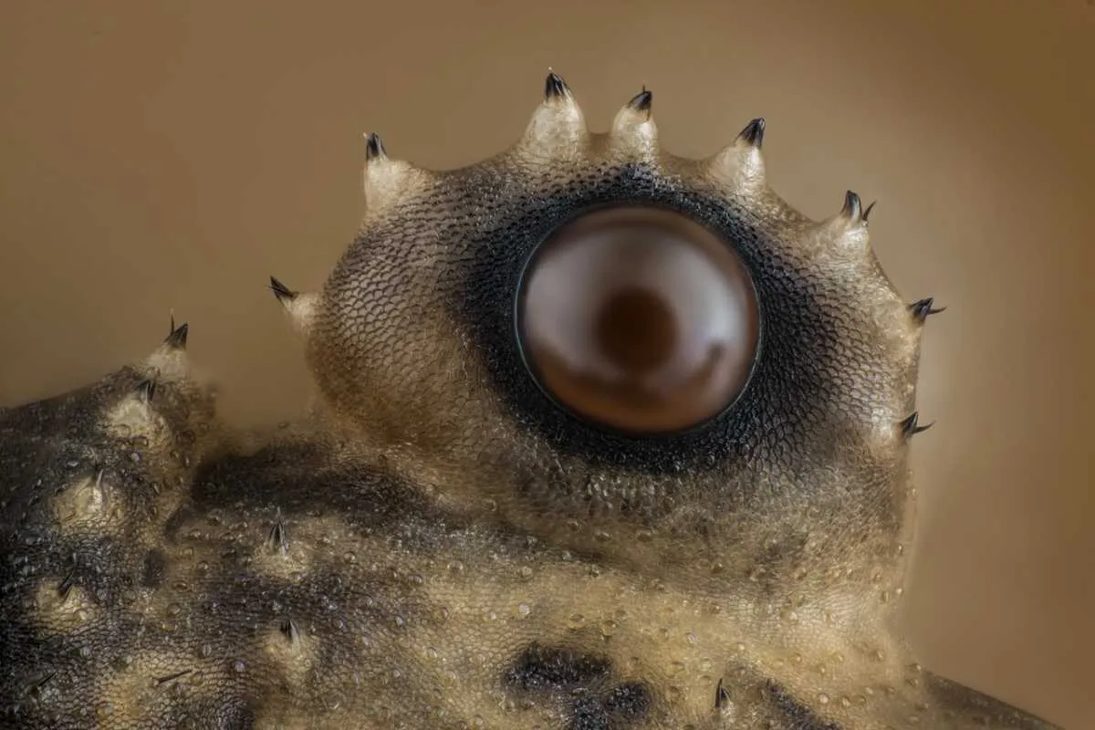
12th Place - Charles Krebs
Charles Krebs Photography
Issaquah, Washington, USA
Opiliones (daddy longlegs) eye
Reflected Light, Image Stacking, 20x (objective lens magnification)
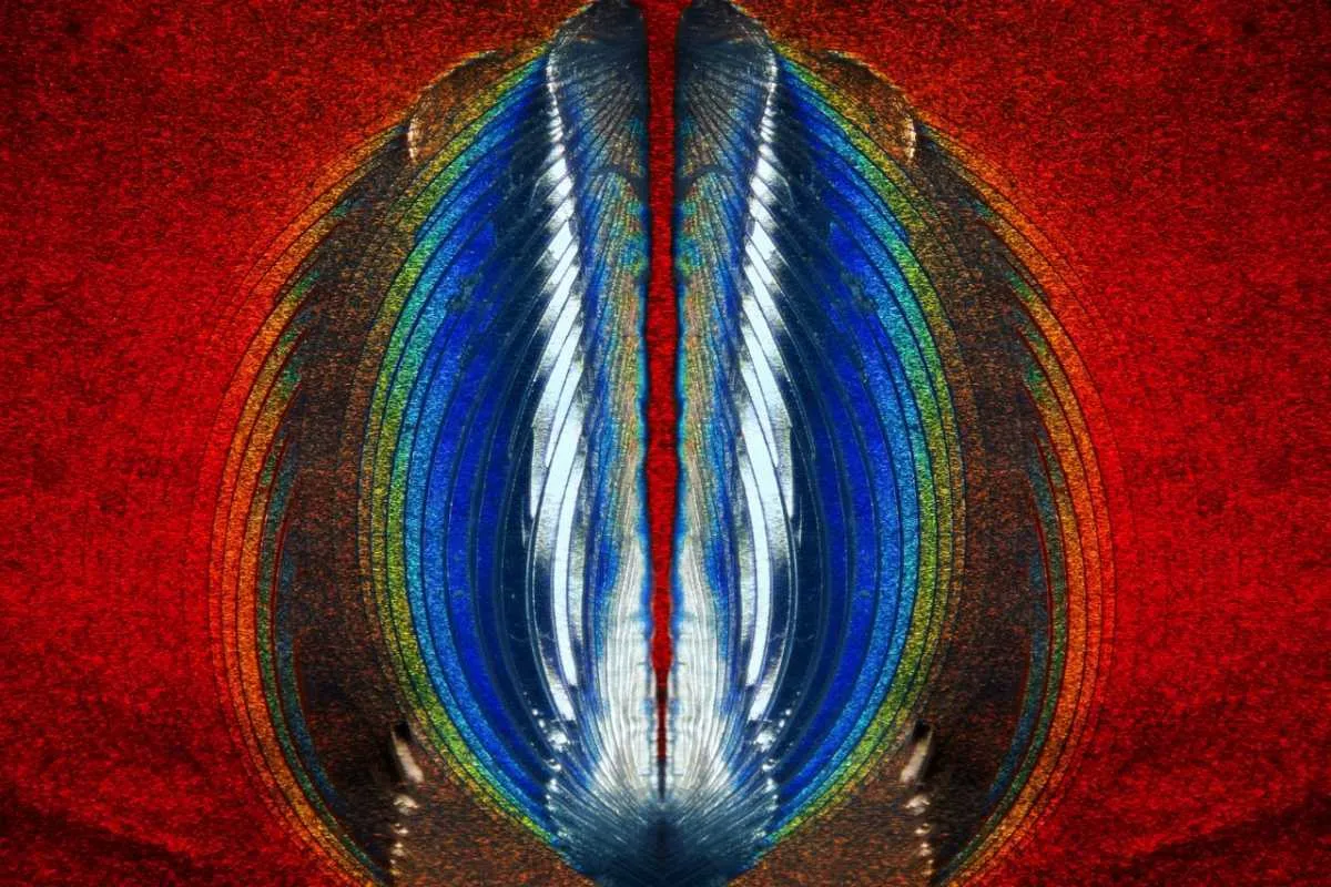
11th Place - Steven Simon
Simon Photography
Grand Prairie, Texas, USA
Plastic fracturing on credit card hologram
10x (objective lens magnification)
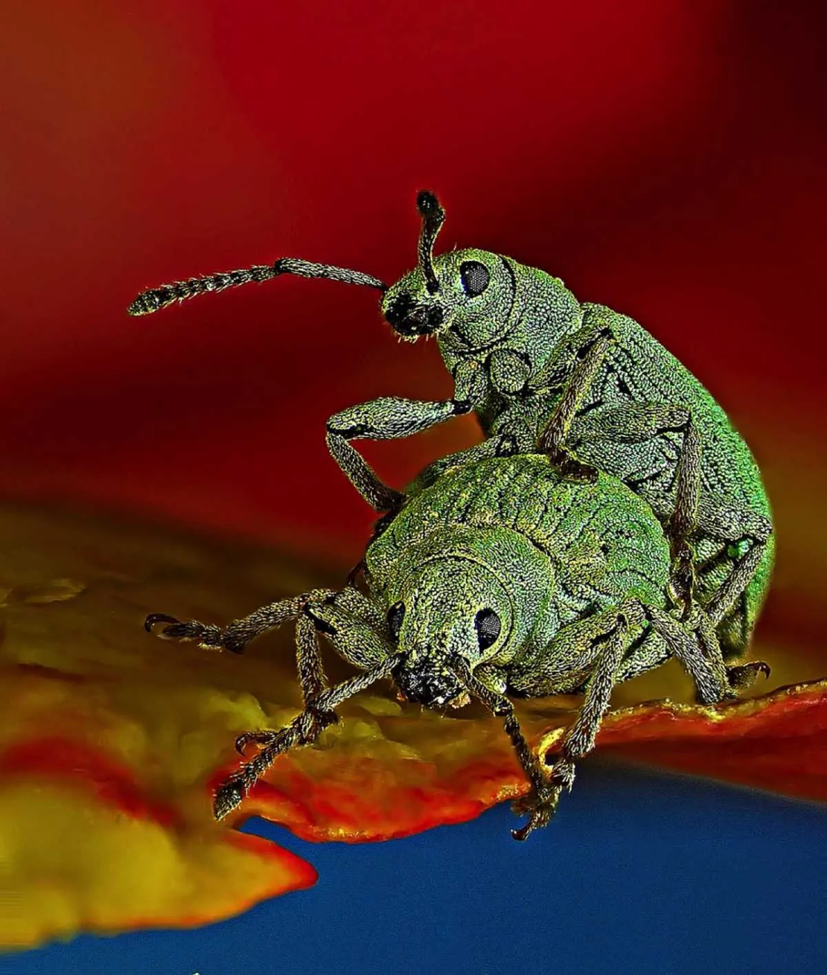
10th Place - Dr. Csaba Pintér
University of Pannonia, Georgikon Faculty, Department of Plant Protection
Keszthely, Hungary
Phyllobius roboretanus (weevil)
Stereomicroscopy, 80x
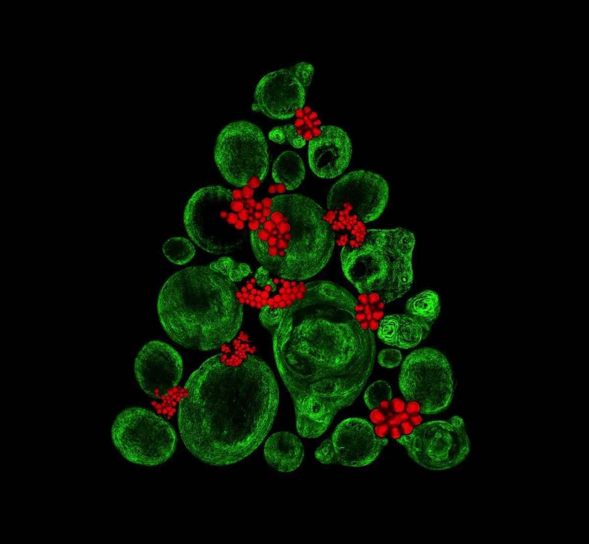
9th Place - Catarina Moura, Dr. Sumeet Mahajan, Dr. Richard Oreffo & Dr. Rahul Tare
University of Southampton, Institute for Life Sciences
Southampton, United Kingdom
Growing cartilage-like tissue in the lab using bone stem cells (collagen fibers in green and fat deposits in red)
Second Harmonic Generation (SHG) and Coherent Anti-Stokes Raman Scattering (CARS), 20x for collagen; 40x for fat deposits
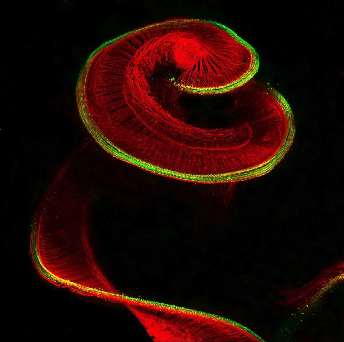
8th Place - Dr. Michael Perny
University of Bern, Institute for Infectious Diseases
Bern, Switzerland
Newborn rat cochlea with sensory hair cells (green) and spiral ganglion neurons (red)
Confocal, 100x
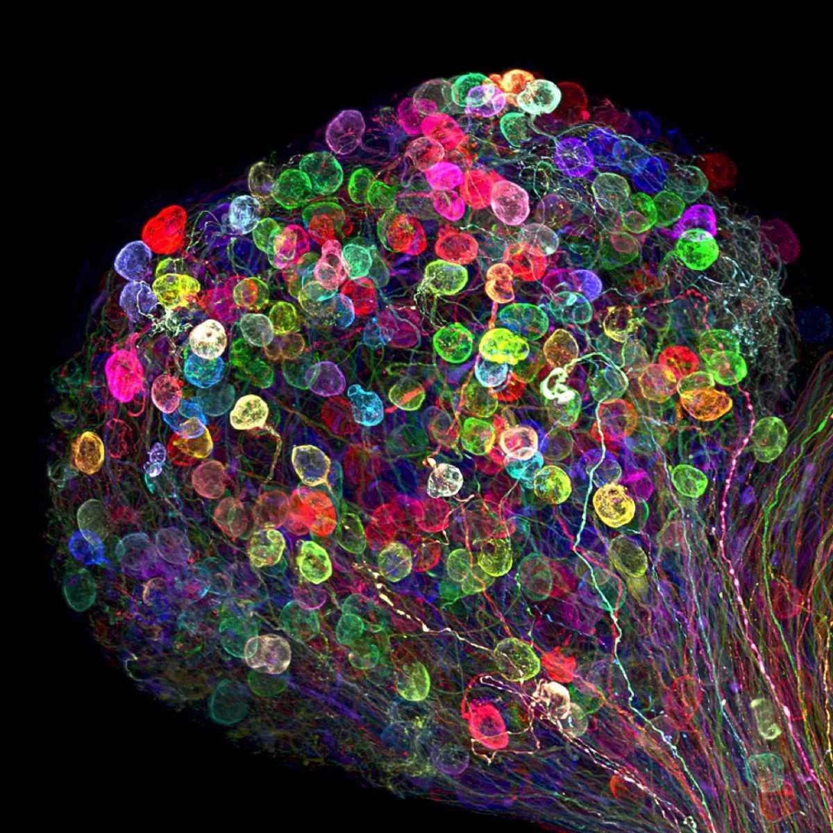
7th Place - Dr. Ryo Egawa
Nagoya University, Graduate School of Medicine
Nagoya, Japan
Individually labelled axons in an embryonic chick ciliary ganglion
Differential Interference Contrast
Confocal, Tissue Clearing, Brainbow (labeling technique), 30x (objective lens magnification)
6th Place - Dr. David A. Johnston
University of Southampton/University Hospital Southampton, Biomedical Imaging Unit
Southampton, United Kingdom
Lily pollen
Confocal, 63x (objective lens magnification)
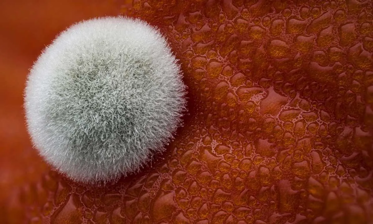
5th Place - Dean Lerman
Netanya, Israel
Mold on a tomato
Reflected Light, Focus Stacking, 3.9x
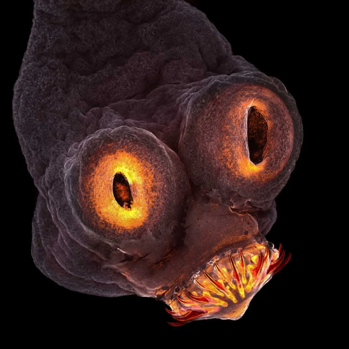
4th Place - Teresa Zgoda
Rochester Institute of Technology
Rochester, New York, USA
Taenia solium (tapeworm) everted scolex
200x
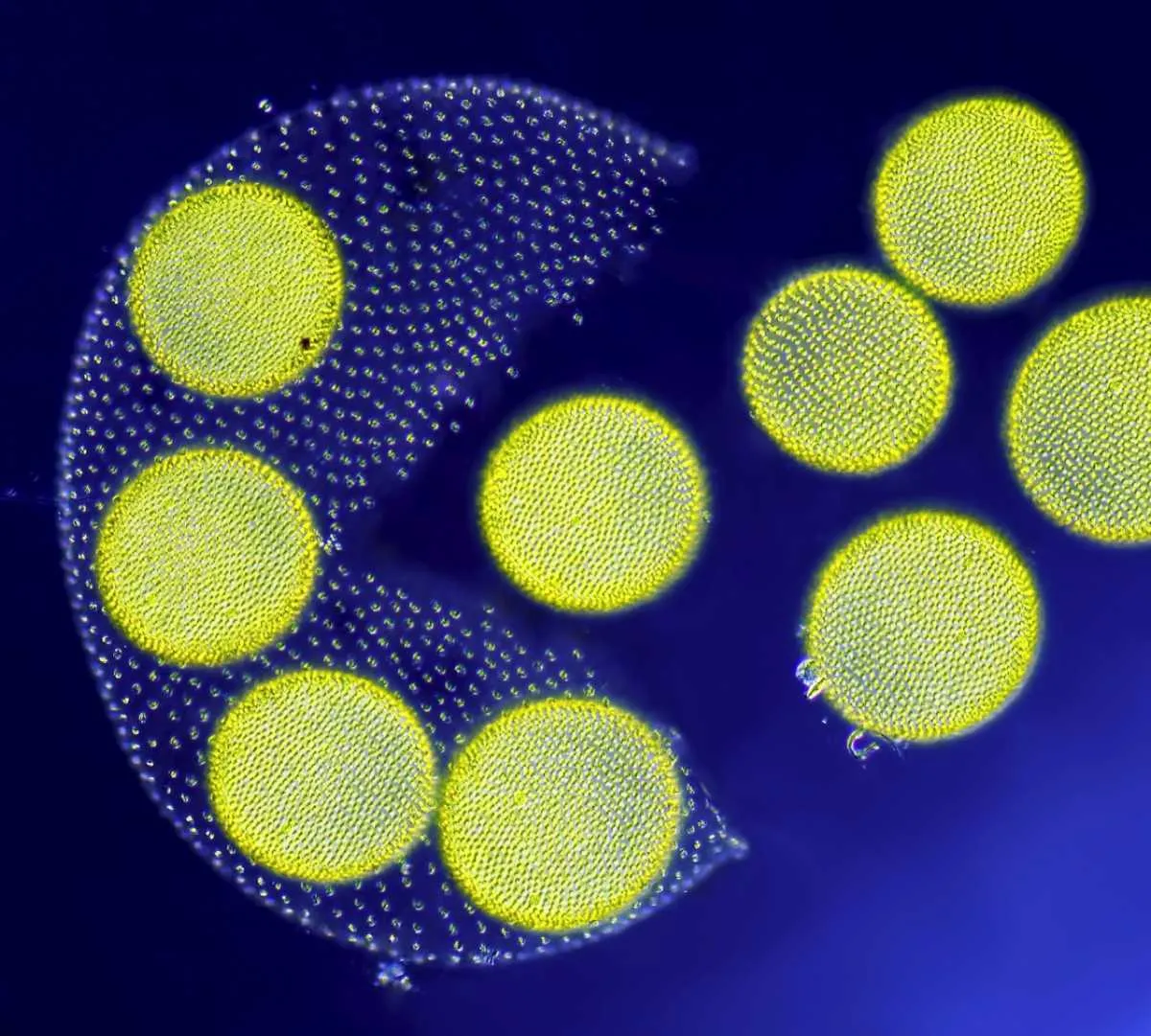
3rd Place - Jean-Marc Babalian
Nantes, France
Living Volvox algae releasing its daughter colonies
Differential Interference Contrast, 100x

2nd Place - Dr. Havi Sarfaty
Eyecare Clinic
Yahud-Monoson, Israel
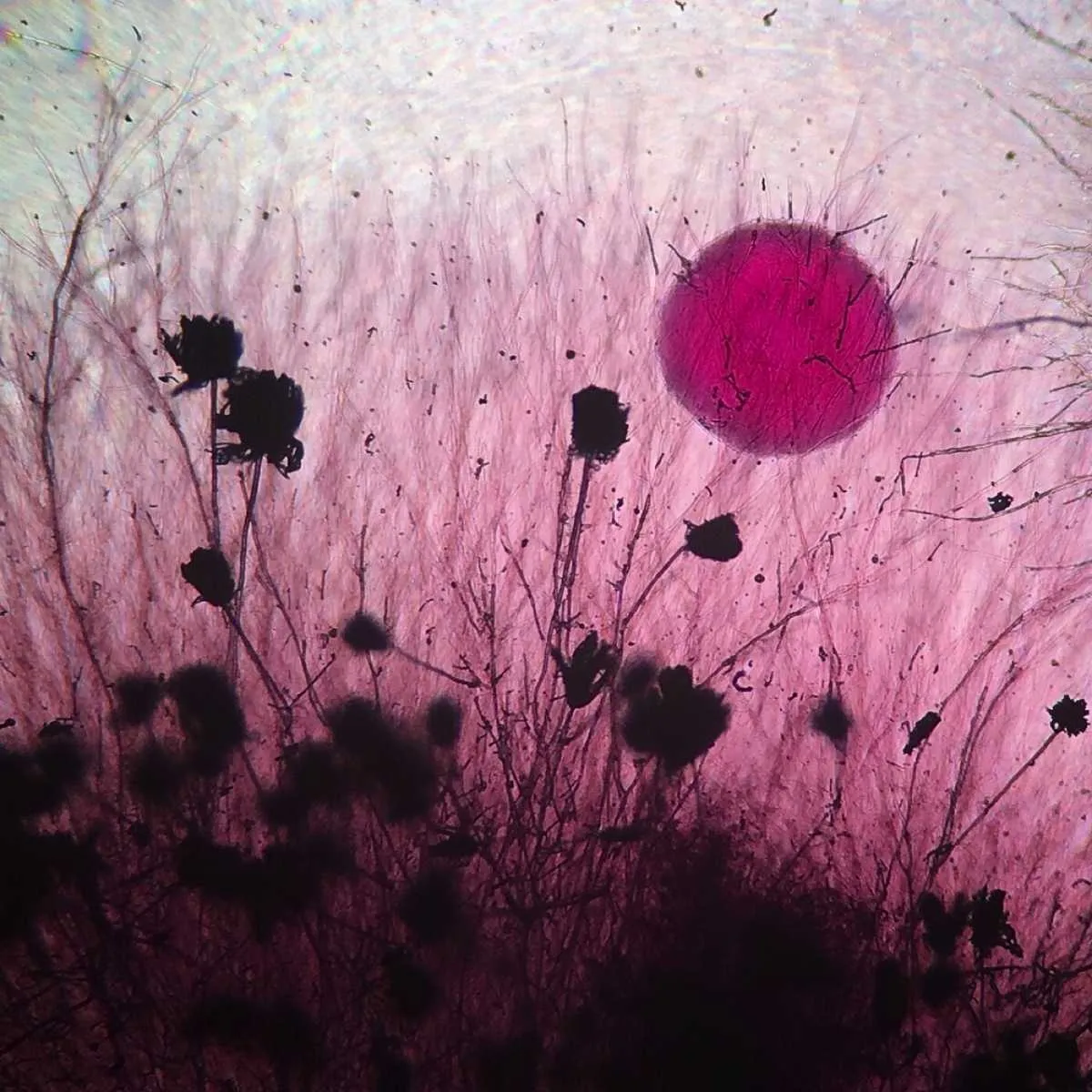
Senecio vulgaris (a flowering plant) seed head
Stereomicroscopy, 2x
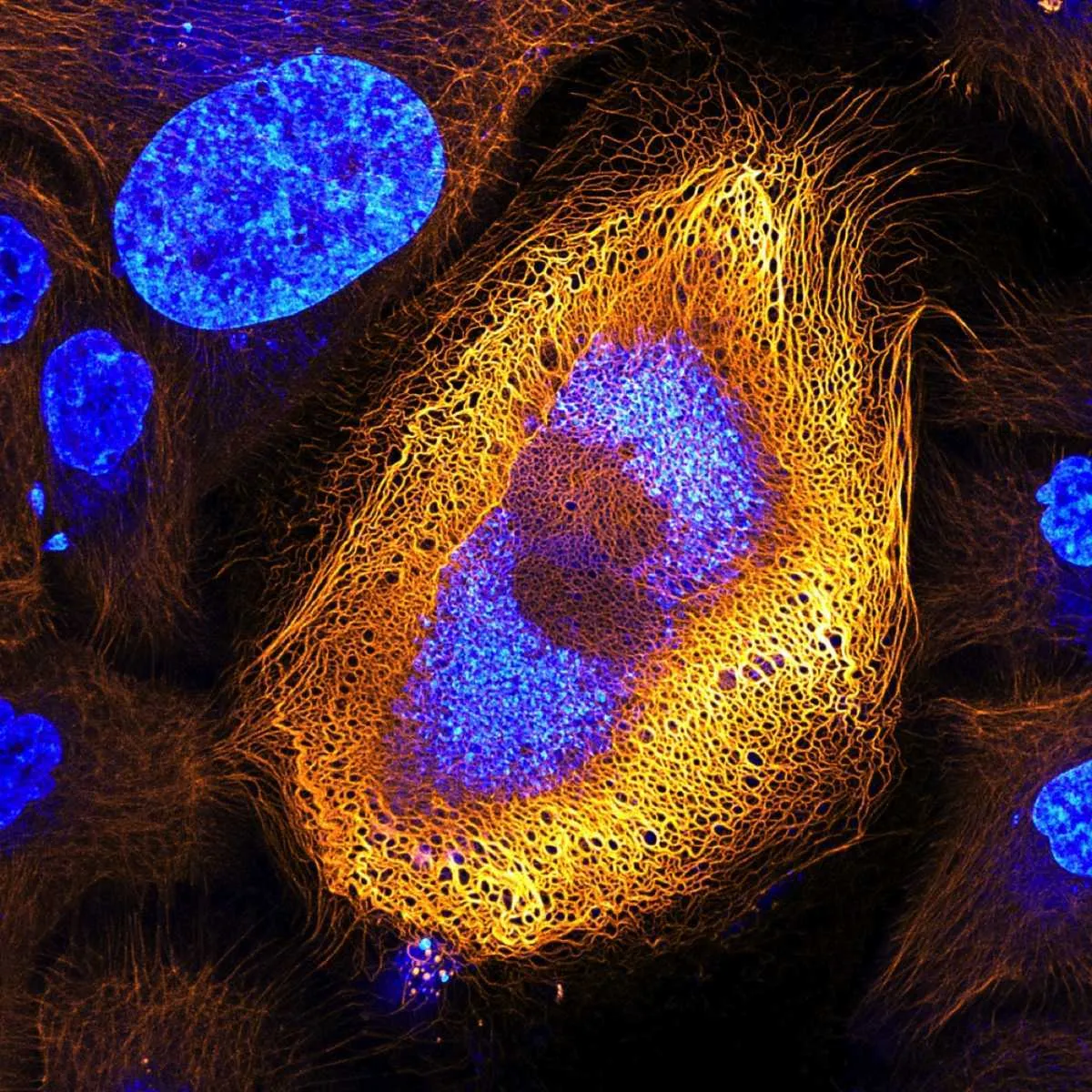
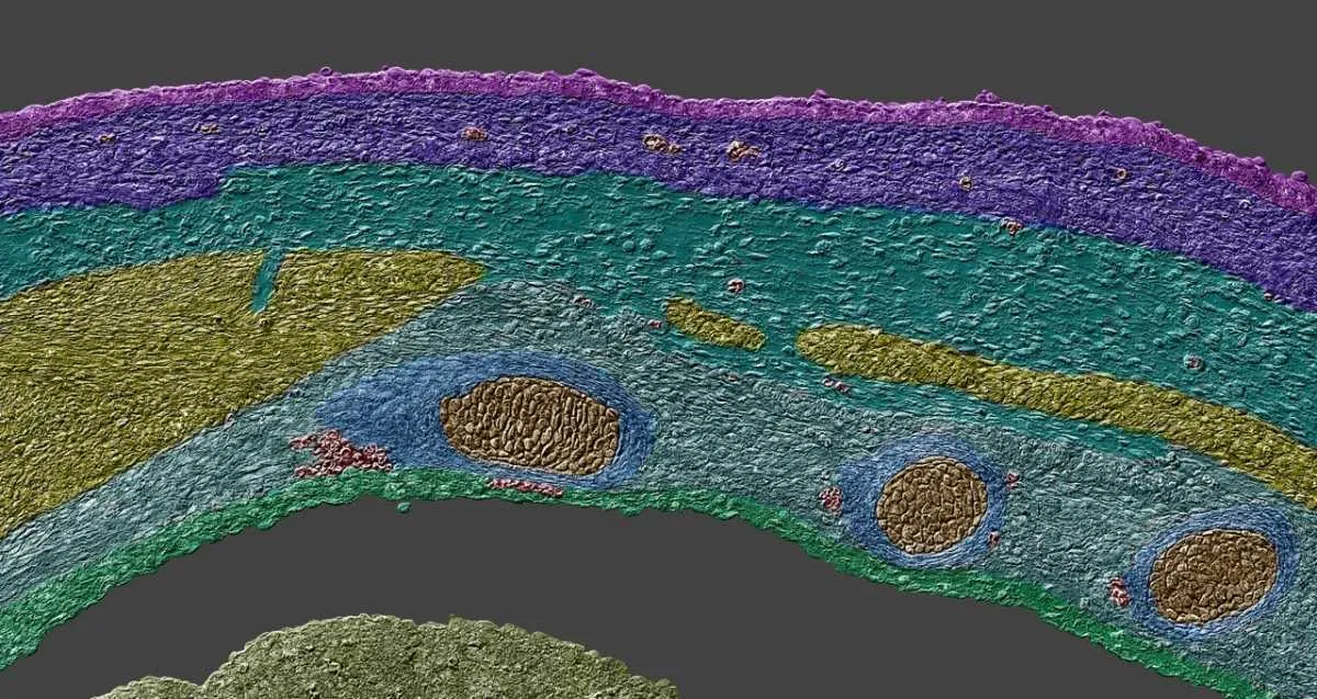
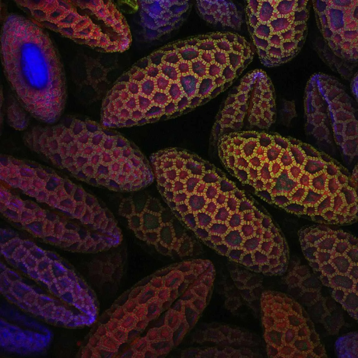
1st Place - Dr. Bram van den Broek, Andriy Volkov, Dr. Kees Jalink, Dr. Nicole Schwarz & Dr. Reinhard Windoffer
The Netherlands Cancer Institute, BioImaging Facility & Department of Cell Biology
Amsterdam, The Netherlands
Immortalized human skin cells (HaCaT keratinocytes) expressing fluorescently tagged keratin
Confocal, 40x (objective lens magnification)
Follow Science Focus onTwitter,Facebook, Instagramand Flipboard

