We spend our entire lifetime encased in our bodies and you might believe you have an intimate knowledge of yourself. If invited to, you could conjure up a reasonably accurate mental image of your face or figure. But how much of you do you know?
In a new book from Bonnier and the Wellcome Collection, curators Dr Jennifer Z Paxton and Katy Wiedemann take the reader on a journey underneath the skin. An exploration of the human body, Anatomicum is a museum quite unlike any other – open 24 hours a day, seven days a week, this detailed look at our inner workings will remind you just how ingenious every part of us is.
The extracts below have been chosen by Jennifer, who wrote the text, and Katy, who provided stunningly detailed artwork.
The Skeletal System
Providing the framework for the entire human body is the skeleton. This hard but flexible scaffold gives us our overall shape, supports the muscles, protects the body’s soft inner organs and constantly manufactures new blood cells. The entire skeleton is
made up of 206 individual bones, which link together at areas called joints. These keep the bones of the skeleton fixed to each other, while also allowing us to move.
Anatomists split the skeleton into two parts: the axial skeleton, which supports the upper body and protects the inner organs; and the appendicular skeleton, made up of the bones in the limbs and pelvis. The axial skeleton incorporates all the bones of the head and trunk, where its primary role is to protect the brain, the sensory organs (eyes, ears and nose), the heart and the lungs. It also includes the vertebral column, or spine, a long row of 33 bones called vertebrae which encase the delicate spinal cord. Stacked together, the vertebrae make a curved, flexible pillar, which can bend forwards, backwards or side-to-side. The spine connects to the skull at its first (or top) vertebra, which is named after the figure Atlas from ancient Greek mythology. Atlas was condemned to bear the weight of the whole sky on his shoulders, just as the atlas vertebra has to support the weight of the skull and brain.
The bones of the upper limbs – the arm, forearm, wrist and hand – have a greater range of movement than any other part of the skeleton, combining strength with dexterity so that we can perform tasks as varied as threading a needle and batting a ball. In contrast, the bones of the lower limbs – in the thigh, leg, ankle and foot – mainly provide stability and support our weight as we walk, stand or run. Key to this is the longest and strongest bone in the human body: the femur or thigh-bone.
Key to plate
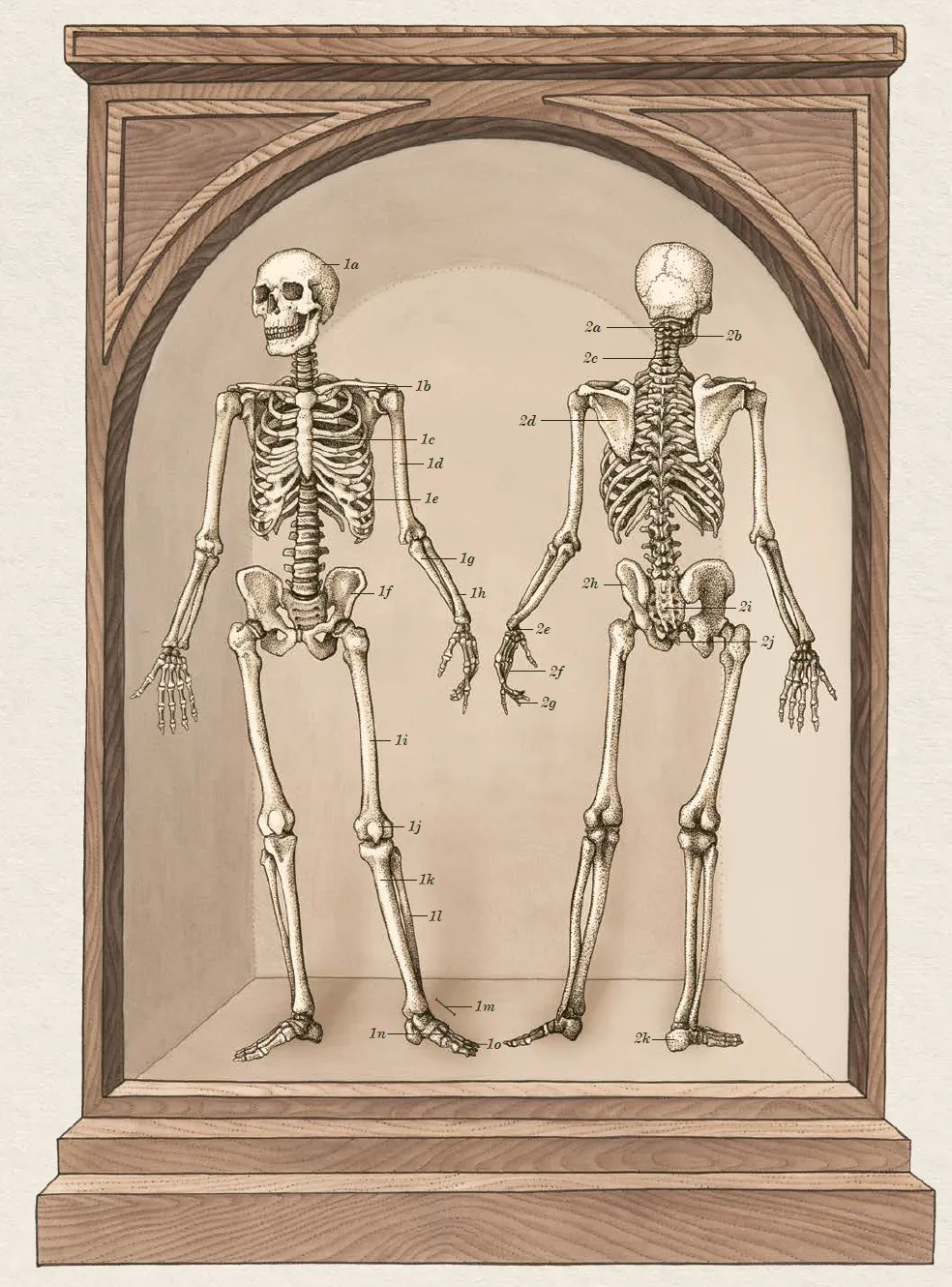
1. Skeleton, seen from the front (anterior view)
- a) Cranium (skull bones)
- b) Clavicle (collar bone)
- c) Sternum (breast bone)
- d) Humerus (arm bone)
- e) Ribs
- f) Pelvis (hip bones)
- g) Ulna (inner forearm bone)
- h) Radius (outer forearm bone)
- i) Femur (thigh bone)
- j) Patella (knee cap)
- k) Tibia (inner leg bone)
- l) Fibula (outer leg bone)
- m) Tarsal bones (ankle and foot bones)
- n) Metatarsals (bones of the foot)
- o) Phalanges (toe bones)
2. Skeleton, seen from the back (posterior view)
- a) Atlas (1st spine bone)
- b) Axis (2nd spine bone)
- c) Vertebral column (spine or backbone)
- d) Scapula (shoulder blade)
- e) Carpal bones (wrist bones)
- f) Metacarpals (hand bones)
- g) Phalanges (finger bones)
- h) Ilium (one of the pelvis bones)
- i) Sacrum (fused bones at base of spine)
- j) Coccyx (tail bone)
- k) Calcaneus (heel bone)
The Skull
Underneath the skin and muscles of our head lies the skull, a protective home for the brain and sensory organs (the eyes, ears, nose and tongue). Although it appears to be a single bone, it is in fact formed of 22 individual bones, most of which are fused together at non-moveable joints known as sutures. The top part, or vault, is formed of eight bones and acts like a helmet, shielding the delicate brain inside from injury. The other 14 bones provide shape for the face and jaw. Only one of these, the mandible or jaw-bone, can move. This bone is joined to the skull by a hinge joint, letting us open and close the jaw during chewing and talking.
Most of the bones in the face are not solid, but have air-filled spaces within them called sinuses. These reduce the overall weight of the skull and add a deeper, clearer sound to our voices during speech, by allowing air to vibrate within them. In addition to sinuses, there are a number of holes running right through the skull bones, known as foramina. These let the brain connect with other parts of the body via nerves, and allow blood vessels to pass to and from the brain and face. The largest hole in the entire skull is at its base and is the foramen magnum, meaning ‘great hole’. This large, oval shaped space is where the brain joins to the spinal cord.
You might notice that some facial features are missing from the skull – there are no ears or nose visible. This is because the shape of the outer nose and ear is made from cartilage, a material which is softer, and decays more rapidly, than bone.
After death, a skull may provide many clues about who it belonged to. We can ascertain age, gender and ethnicity by studying the skull’s size and features, and (as with other bones) it is possible to see what diseases a person may have suffered from or even where they may have lived. Scientists known as palaeoanthropologists can study bones to discover facts about ancient cultures, while forensic osteologists study bones to uncover vital clues about the cause of death in criminal cases.
Key to plate
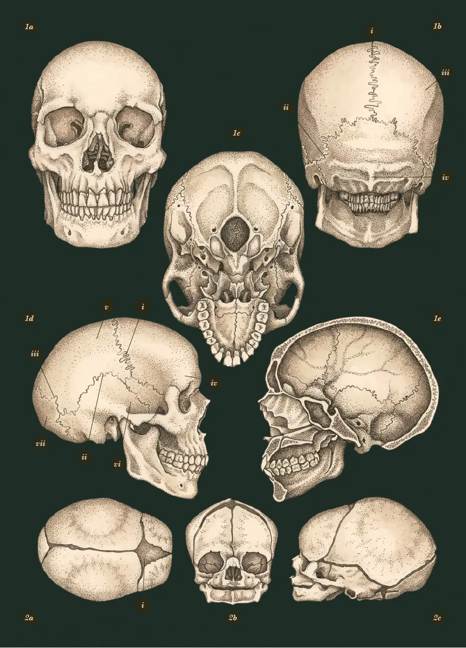
1: Adult skull
- a) From the front
- b) From the back: The sagittal (i) and lambdoid (ii) sutures can be seen, that join the parietal (iii) and occipital (iv) skull bones together.
- c) From the base (without jaw): The large central hole in the base of the skull is called the foramen magnum. This is the point where the spinal cord passes out of the skull to travel down the vertebral column.
- d) From the side: The coronal (i), squamous (ii) and lambdoid (iii) sutures, which join the frontal (iv), parietal (v), temporal (vi) and occipital (vii) skull bones together.
- e) Cross-section from the side: The space inside the skull where the brain sits is called the cranial fossa.
2: Newborn skull
Although the bones of an adult skull are fused together and cannot move, in babies, these joints have not yet hardened. Instead, the sutures are formed from a much more flexible material, creating ‘soft spots’ or fontanelles in a baby’s skull.
Babies’ brains grow rapidly as they develop, from only about 350g at birth to nearly 600g in the first three months – about half the size of an adult brain. Fontanelles mean that the skull can easily cope with this rapid growth.
- a) From the top: The diamond-shaped area is known as the fontanelle (i), or ‘soft-spot’ of a baby’s skull.
- b) From the front: Infant skulls display more prominent forehead, orbits and a smaller jaw than the typical adult skull.
- c) From the side: The prominent forehead and shape of the small jaw is noticeable.
The Lungs
We breathe over 10,000 litres of air each day to keep our cells alive and healthy. Getting all of this air in and out of the body is the responsibility of the lungs. These paired organs inhale and exhale air at a rate of around 15 breaths a minute, and transfer oxygen from inhaled air into the bloodstream, while passing carbon dioxide from the bloodstream into the air to be exhaled.
Once air enters the lungs, it begins a journey through a dividing network of more tubes, that become smaller and smaller as they travel deeper into the organ. Eventually, the air reaches tiny closed sacs called alveoli, which resemble small bunches of grapes. Each individual alveolus (singular of alveoli) is covered in a tiny blood vessel called a capillary. The walls of the alveoli and the capillaries are only one cell layer thick (less than a thousandth of a millimetre), so gases can easily be exchanged to and from the blood. There are several hundred million alveoli in an adult’s lungs, meaning they have a huge surface area in order to quickly transfer as much gas as possible. In fact, the total surface area of the lungs is estimated to be bigger than that of a tennis court!
The right and left lungs sit within the chest, under the ribcage. The right lung has three different parts, or lobes, and is a little bigger than the left, which only has two lobes. The left lung is smaller to make room for the heart that fits snugly between the two lungs. A large, dome-shaped muscle called the diaphragm is found directly underneath the lungs, separating them from the organs in the abdomen. The diaphragm helps to create space in the chest when we breathe in, or inhale, by pulling downwards and flattening out. This lets the lungs fill with air, inflating like balloons and raising the ribcage outwards. When we breathe out, or exhale, the diaphragm relaxes and pushes on the lungs, squeezing the air back out again. This cycle happens between 12 and 20 times each minute when resting and much more often during exercise as breathing rate increases.
Key to plate
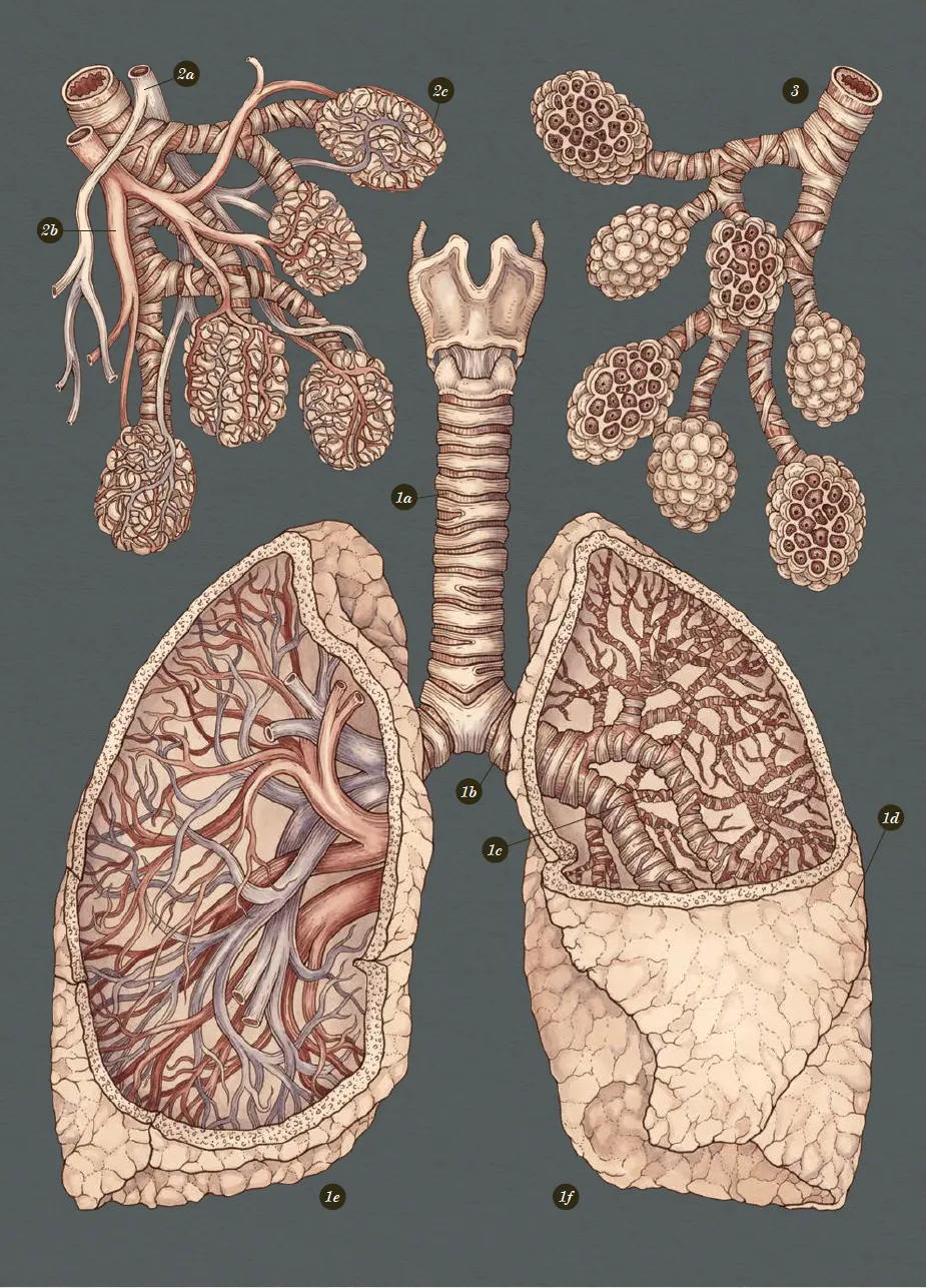
1: Lungs
a) Trachea
b) Bronchi
c) Bronchioles
d) Pleural membranes: Two thin, fluid-filled membranes encase the lungs. They enable them to move smoothly as the lungs inflate and deflate.
e) Right lung
f) Left lung
2: Alveoli
a) Venule
b) Arteriole
c) Capillary bed
The millions of alveoli in each lung group together to form small bunches. Tiny capillaries wrap around them so that gas can easily transfer between the blood and the air in the alveoli.
3: Cross-section of alveoli
Inside each grapelike cluster are dozens of individual alveoli. Each one has a wall just one cell-layer thick, so oxygen can enter the bloodstream as easily as possible.
Anatomicum by Jennifer Z Paxton, Katy Wiedemann is available now (£25, Wellcome)
Follow Science Focus onTwitter,Facebook, Instagramand Flipboard