Instead of bucolic landscapes, deep-and-meaningful portraits, and hipster street photography, the winners of the Nikon Small World Photomicrography Competition turn their cameras on the microscopic world, which means this year’s collection is filled with snowflakes, spiders and see-through embryos.
Now in its 45th year, the competition has celebrated scientific images and the technology behind them, and has produced an amazing array that are as enthralling as they are beautiful (see 2017’s competition for a delightful algae that looks like the video game character Pac-Man).
So without further ado, here are the top 20 winners from the Nikon Small World Photomicrography Competition 2019.
1
Fluorescent turtle embryo
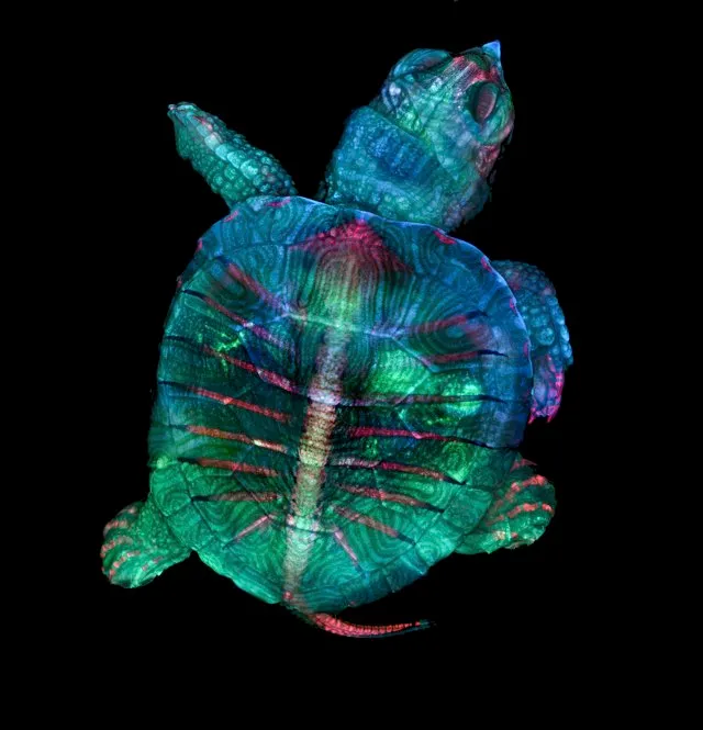
Teresa Zgoda & Teresa Kugler
Campbell Hall, New York, USA
Stereomicroscopy, Fluorescence
5x (Objective Lens Magnification)
2
Depth-colour coded projections of three stentors (single-cell freshwater protozoans)
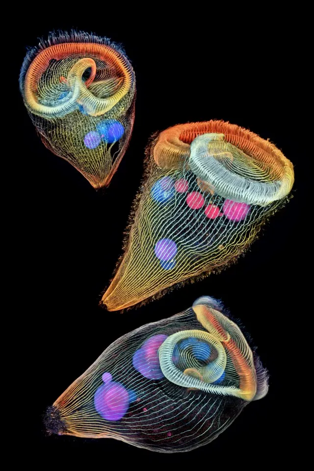
Dr Igor Siwanowicz
Howard Hughes Medical Institute (HHMI), Janelia Research Campus, Ashburn, Virginia, USA
Confocal
40x (Objective Lens Magnification)
3
Alligator embryo developing nerves and skeleton
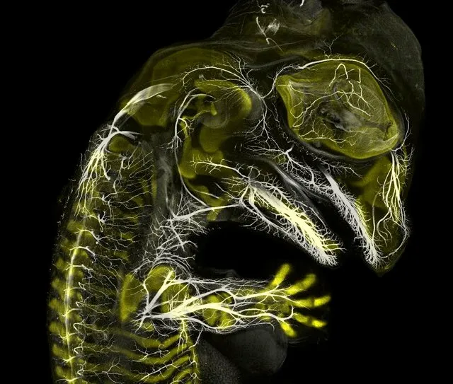
Daniel Smith Paredes & Dr. Bhart-Anjan S. Bhullar
Yale University, Department of Geology and Geophysics, New Haven, Connecticut, USA
Immunofluorescence
10x (Objective Lens Magnification)
4
Male mosquito

Jan Rosenboom
Universität Rostock, Rostock, Mecklenburg Vorpommern, Germany
Focus Stacking
6.3x (Objective Lens Magnification)
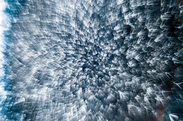
5
Snowflake
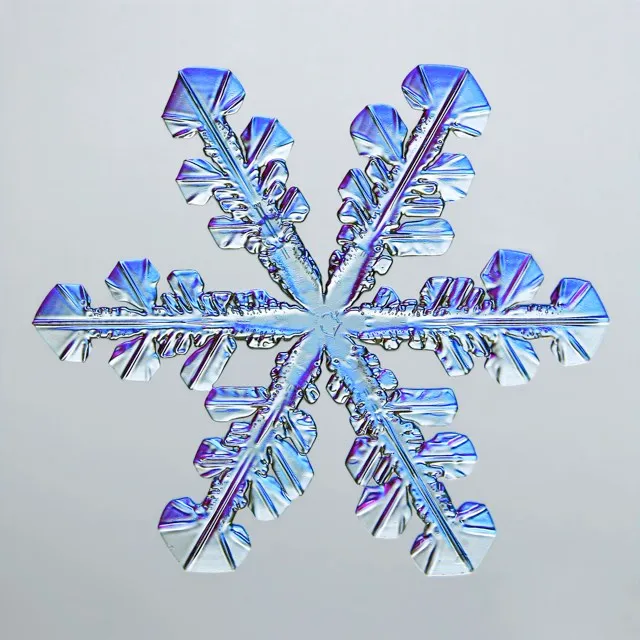
Caleb Foster
Caleb Foster Photography, Jericho, Vermont, USA
Transmitted Light
4x (Objective Lens Magnification)
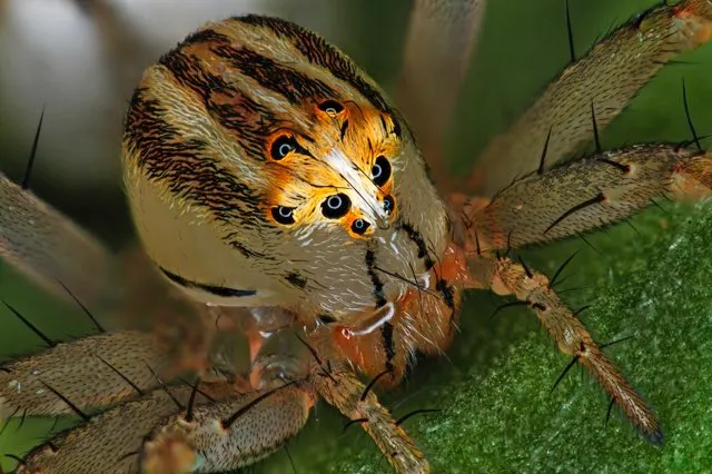
6
Small white hair spider
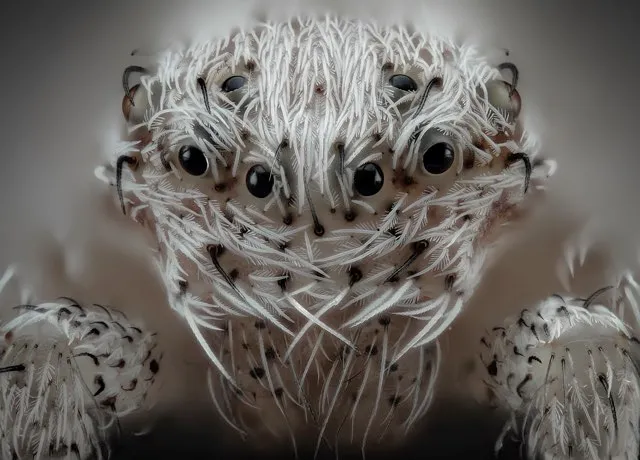
Javier Rupérez
Almáchar, Málaga, Spain
Reflected Light, Image Stacking
20x (Objective Lens Magnification)
7
Chinese red carnation stamen

Dr. Guillermo López López
Alicante, Spain
Focus Stacking
3x (Objective Lens Magnification)
8
Frozen water droplet
Garzon Christian
Quintin, Cotes-d’Armor, France
Incident Light
8x (Objective Lens Magnification)
9
Tulip bud cross section
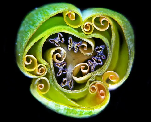
Andrei Savitsky
Cherkassy, Ukraine
Reflected Light
1x (Objective Lens Magnification)
10
BPAE cells in telophase stage of mitosis
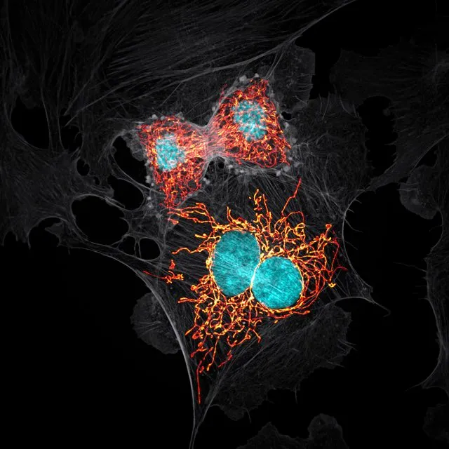
Jason M. Kirk
Baylor College of Medicine, Optical Imaging & Vital Microscopy Core,Houston, Texas, USA
Confocal with Enhanced Resolution
63x (Objective Lens Magnification)
11
A pair of ovaries from an adult Drosophila female stained for F-actin (yellow) and nuclei (green); follicle cells are marked by GFP (magenta)
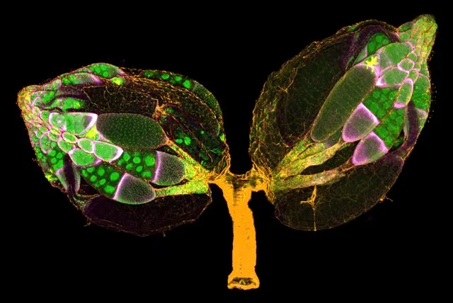
Dr. Yujun Chen & Dr. Jocelyn McDonald
Kansas State University,Department of Biology, Manhattan, Kansas, USA
Confocal
10x (Objective Lens Magnification)
12
Mosquito larva
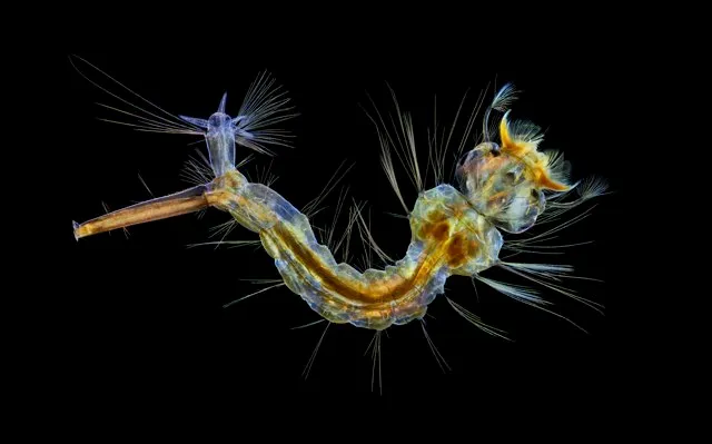
Anne Algar
Hounslow, Middlesex, United Kingdom
Darkfield, Polarizing Light, Image Stacking
4x (Objective Lens Magnification)
13
Cuprite (mineral composed of copper oxide)
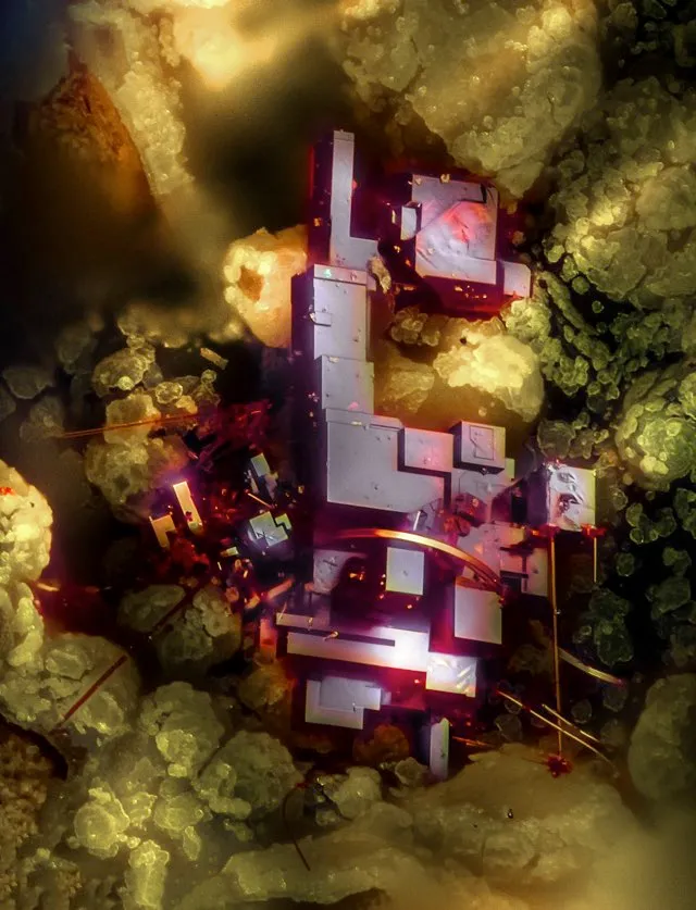
Dr. Emilio Carabajal Márquez
Madrid, Spain
Focus Stacking
20x (Objective Lens Magnification)
14
Female Oxyopes dumonti (lynx) spider
Antoine Franck
CIRAD - Agricultural Research for Development,Saint Pierre, Réunion
Focus Stacking
1x (Objective Lens Magnification)
15
Pregnant Daphnia magna (small planktonic crustacean)
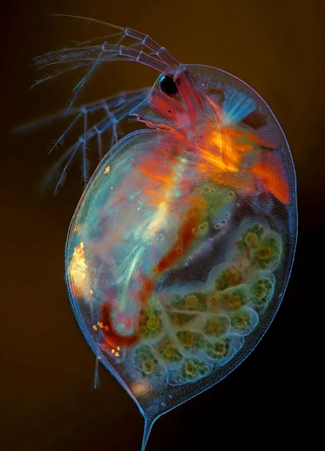
Marek Miś
Marek Miś Photography,Suwalki, Podlaskie, Poland
Modified Darkfield, Polarized Light, Image Stacking
4x (Objective Lens Magnification)
16
Housefly compound eye pattern
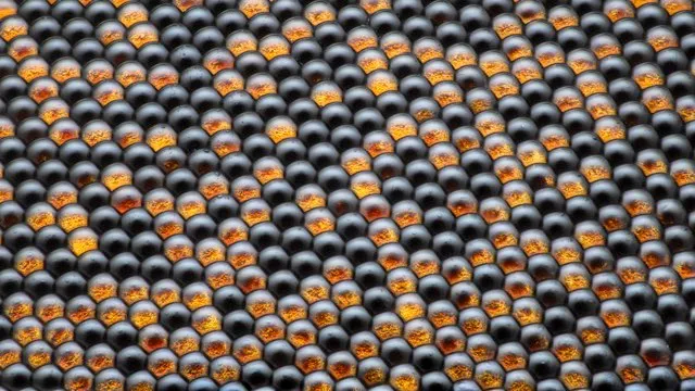
Dr. Razvan Cornel Constantin
Bucharest, Romania
Focus Stacking, Reflected Light
50x (Objective Lens Magnification)
17
Vitamin C
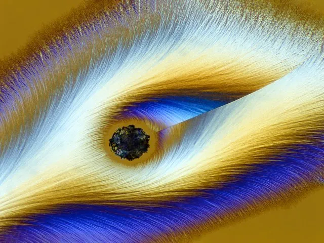
Karl Deckart
Eckental, Bavaria, Germany
Brightfield, Polarized Light
4x (Objective Lens Magnification)
18
Cristobalite crystal suspended in its quartz mineral host
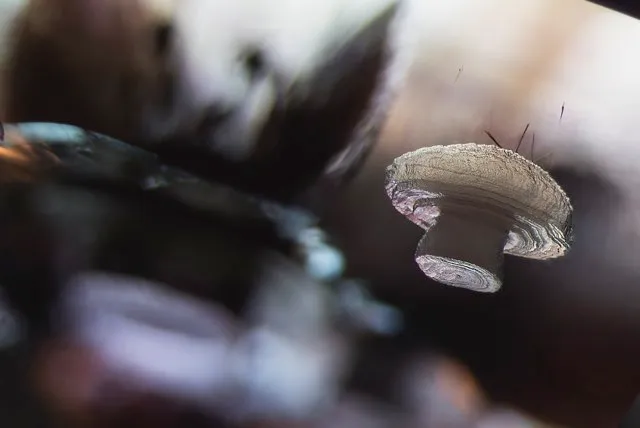
Billie Hughes
Lotus Gemology,Bangkok, Thailand
Darkfield
40x (Objective Lens Magnification)
19
Octopus bimaculoides embryo
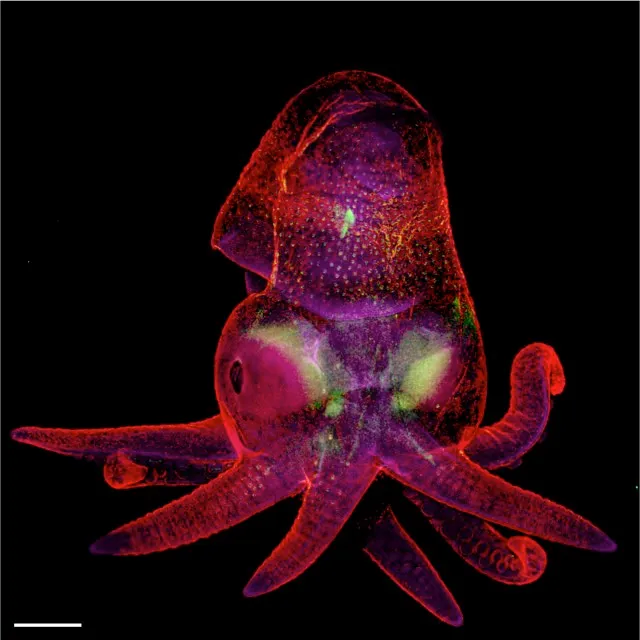
Martyna Lukoseviciute & Dr. Carrie Albertin
University of Oxford, Weatherall Institute of Molecular Medicine,Oxford, Oxfordshire, United Kingdom
Confocal, Image Stitching
5x (Objective Lens Magnification)
20
Blood vessels of a murine (mouse) heart following myocardial infarction (heart attack)
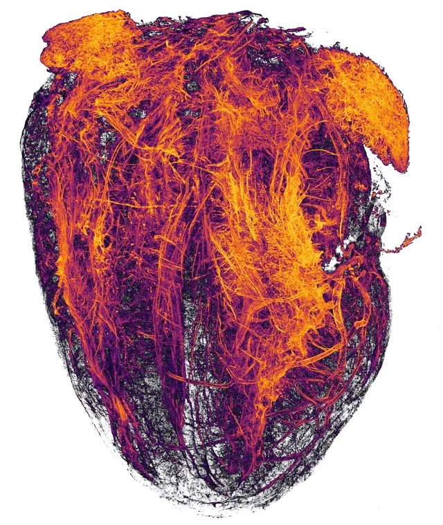
Simon Merz, Lea Bornemann & Sebastian Korste
University Hospital Essen,Institute for Experimental Immunology & Imaging, Essen, Nordrhein-Westfalen, Germany
Tissue Clearing, Light Sheet Fluorescence Microscopy
2x (Objective Lens Magnification)